


Osteochondrosis in the lumbar region is a chronic degenerative disease of the lumbar column which affects the structure of intervertebral discs and a number of lumbar vertebrae located.He affects people of predominantly.It manifests in various symptoms, the main ones of which are pain in the lower back and legs, limiting movements in the lower back.For diagnoses, research methods such as radiography, computed tomography or magnetic resonance tomography of the lumbar column are used.In this article, you can in more detail with the causes, symptoms and methods for diagnosing osteochondrosis in the lumbar column.
Osteochondrosis is the result of aging the body.These signs or others of this disease are in almost everyone (!), From 25 years old.But here is the severity of these changes, the rate of their progression, the degree of clinical manifestations depends on many causes, mainly how a healthy lifestyle leads to a specific person.Moderate physical activity, compulsory morning gymnastics, the good installation of the body when carrying out a certain number of works (garden, construction, commonplace cleaning of the house, etc.), the orthopedic mattress is the moments that prevent the development of osteochondrosis of the lumbar spine.
According to statistics, osteochondrosis of the spine in 80% of cases is the cause of back pain.
How is osteochondrosis develop?
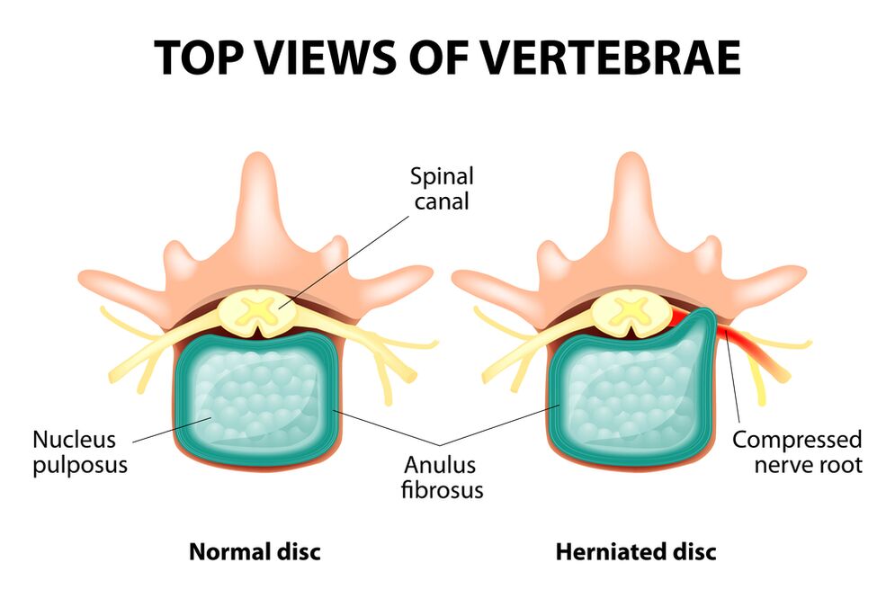
The whole spine consists of separate vertebrae, between the bodies of which there are intervertebral discs.In other words, between the two vertebrae is a disc.The disc consists of a gelatinous (luscious) nucleus and a fibrous ring.The nucleus contains a lot of water and offers depreciation and flexibility of the spine.The fibrous ring is located along the periphery of the nucleus of the jacket, as if it kept it inside itself.
With an increased increased load on the core of the pier, it modifies its physiological properties, loses water and dry, and possibly sequences: the disc is flattened and vertebral bodies are closer to each other.With such processes, in the nucleus jacket, the fibrous ring loses its elasticity and, under the influence of mechanical loads, begins to overcome.This is called protrusion.Then, the fibrous ring cracks and a gelatin nucleus falls through the resulting gaps: a hernia of the disc occurs.A plot of two adjacent vertebrae and a disc located between them, called the vertebral segment, acquires excessive mobility, thus increasing the load on the neighboring segments.The overload of neighboring segments triggers a similar pathological process.These changes are called osteochondrosis.
In order to ensure the stability of the spine, bone growths are formed along the edges of vertebral bodies, increasing the area of support.This phenomenon is called spondylosis.The changes in the joints between the vertebrae are called spondylo osteoarthritis.Usually, the three pathologies - osteochondrosis, spondylosis, spondyl osteoarthritis - walk nearby.
Reasons
Why does osteochondrosis occur?To date, there are several theories of the event:
- Mechanical theory: perhaps the main reason should be considered as a regular increased load on the spine.This is why osteochondrosis is an almost compulsory destiny of movers, minors, manufacturers and people from such professions.The occurrence of osteochondrosis in the lumbar region is mainly associated with the slopes and the lifting of gravity, forced to install uncomfortable work;
- Another development factor is an incorrect posture, seated in poor pose, which is particularly relevant for mental workers;
- Sometimes the role is played by hereditary characteristics of the structure of the spine and the nutrition of its individual structures;
- Traumatic theory: any trauma of the spine (even the most insignificant) is capable of launching a degenerative process;
- Hormonal metabolic disorders and endocrine diseases can harm metabolism in the spine tissues and contribute to the development of osteochondrosis;
- Age theory implies the natural wear and tear of discs in the life process.
Rarely, only one of these theories can explain the occurrence of osteochondrosis in each case.More often than not, at the same time, several factors are "to blame".
In the occurrence of osteochondrosis of the lumbar column, overweight plays an important role, because it is itself an overload for the spine.The higher the body mass index (degree of obesity), the more changes in changes in the spine are generally pronounced.Among other reasons causing the appearance of osteochondrosis, we can note:
- sedentary lifestyle;
- Imorious nutrition (fast food, excess of sweet and semi-finished products: all this leads to an imbalance in trace elements) and a lack of liquid;
- Anomalies of the structure of the spine (for example, the presence of an additional lumbar vertebra);
- Constant port of high themed shoes;
- pregnancy (due to excess load on the lumbar column);
- Sudden dismissal of training for people professionally involved in sports;
- Smoking and alcohol abuse: as factors that accelerate the aging process in the body.
Symptoms
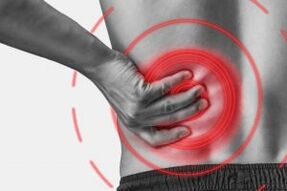
The main manifestation of osteochondrosis in the lumbar column is pain.The nature of the pain, the place of the occurrence and the distribution direction depend on the irritated receptors, that is to say how raw changes in the disc and the surrounding tissues, there is projection or hernia, in which direction the projection was formed and so on.
Reflex and compression syndromes are distinguished by osteochondrosis of the lumbar column.
Reflex syndromes develop in cases where receptors of the fibrous ring of the affected disc, ligaments and articular capsules located nearby are irritated.They are reflexive because in addition to pain is accompanied by changes muscle reflex, vegetative-vascular or neurodistrophic, that is to say that irritation with reflexes is transmitted to other structures, causing symptoms mainly on the side of soft tissues.
Compression syndromes occur following the compression (compression) of the nerve roots, blood vessels or spinal cord formed by osteochondosis by changes.

Lumbar column reflex syndromes
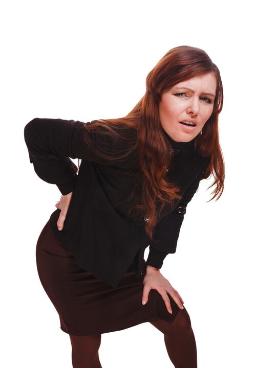
Lumbago(Feeling): Acute sudden pain in the lower back, which occurs with an awkward movement or at the time of physical tension (much less often - for no apparent reason).It is believed that the occurrence of Lumbago is associated with the movement of a nucleus of the jacket in the fibrous ring, that is to say that it develops in the initial stages of osteochondrosis.Often, pain is described as a "feeling", "the stake was stuck in the lower back".Patients freeze in the pose in which pain attracted them.The slightest movement causes an increase in pain (sneezing, coughing, attempted to turn into the bed, to move your foot).If a person was in an inclined position at the time of Lumbago development (which occurs most often), then he cannot straighten up.A muscular tension pronounced in the lumbar column occurs by reflex.Along the vertebrae of this area, a muscle roller is felt, which is sometimes visible to the naked eye without touching, and the muscle tension is so pronounced.Feel painful for the patient.Such an increase in muscle tone plays a immobilizing role, protecting the lumbar segment affected by pathological mobility, which can cause a deterioration of the state.The natural turn of the spine in the lower back (lordosis) is flattened, perhaps the curvature (scoliosis) is possible due to muscle tension.
Low back pain- Another reflex syndrome of the lumbar level.This term also means the presence of pain in the lumbar region.But, unlike Lumbago, the pain does not occur in a sharp way, but gradually, in a few hours, even days.The pain is stupid, moderate intensity, intensifies during the movements, in a sitting or standing position, when it moves from one position to another.A little relief brings the bed or back position with a roll under the lower back, but the passive climb of the straightened leg in this position causes increased pain in the lower back (Lassa symptom).The palpation of the lumbar column is painful, but the reflex tension of the muscles is less pronounced than with Lumbago, and sometimes absent at all.The movements in the lumbar column are limited, but possible.This means that the patient can look and the sides at a certain level (then the pain intensifies).
Sciatica- Another variety of Lumbar level reflex syndrome.By this term, it is pain in the lower back, which gives the buttock and the leg (on the rear surface).The pain is different, mainly painful, but can periodically intensify by the type of "fireplace" in the leg.Just like with low back pain, it intensifies with all movements, walking, constraint, decrease in the bed on the back.The lassa symptom is generally positive.The palpation of the lumbar column is painful, as well as to press certain points (for example, in the middle of the line separating the buttock from the thigh, in the middle of the back of the thigh, in the middle of the popliteal pit).There is a tension of the lower back muscles.The inclinations forward and on the sides are limited.
Lumbar column compression syndromes
The clinical characteristic depends on the structure subject to compression.
Between the vertebrae in each intervertebral hole are nerve roots (spinal nerves): left and right.If the pathological formations for osteochondrosis of the lumbar column (mainly discs of discs) tighten the roots, then radiculopathy develops, whose symptoms differ for each root.The increase in pain during sneezing, cough, movement in the lower back (in particular inclination), the presence of muscle tension in the lower back, the restriction of movements in the lumbar column.The following types of radiculopathies in the lumbar column are the most common:
- Radiculopathy L1, L2, L3: The pain occurs in the lower back, gives the planned thigh.In the same area, the occurrence of paraesthesia (a feeling of crawling cart, numbness) is possible, the superficial sensitivity is disturbed (an acute touch of habit is not distinguished, the feeling of cold and hot) is lost.The knee reflex decreases, the weakness of the quadriceps in the thigh is revealed;
- Radiculopathy L4: The lower back pain gives the front part of the thigh, the inner surface of the knee joint and slightly lower along the inner surface of the lower leg.In the same areas, paresthesia is felt and the surface sensitivity is lost (reduced).The weakness of the quadriceps muscle of the thigh also develops, the knee reflex decreases;
- Radiculopathy L5: one of the frequent locations.The pain gives the buttock, along the outside edge of the thigh, along the front surface of the lower leg to the inside of the foot and the thumb.Paresthesia is felt here, superficial sensitivity is disturbed and pain is given here during sneezing and cough.In addition, there is a difficulty in extending the thumb of the foot, because the muscle that performs this action is innervated by the Kine L5.It is sometimes difficult to stand in the heel with an exposed foot;
- Radiculopathy S1 is also often found with osteochondrosis of the lumbar column.The pain gives the buttock, along the outside edge of the thigh, along the outside edge of the lower leg to the outside edge of the foot and 5th finger, heels.These areas are characterized by a feeling of paresthesia, a decrease in the sensitivity of the surface.The Achilles reflex is reduced.With the damage to this spine, the weakness of the muscles of the lower leg and the flexors of the foot develop, therefore standing and walking on the socks are difficult.
The simultaneous development of radiculopathies of several roots is possible, this is particularly characteristic of L5, S1.It happens that a hernia tightens several roots.
If the record flock decreases, it can squeeze the spinal cord.This is only possible when the hernia is located in the upper reference point, because there are no vertebrae of the spinal cord under the lumbar vertebra II (the roots of the spinal cord are subject to compression, and the ponytail syndrome develops).
If the vessels of the lumbar region are subject to pressures, which perform a blood flow to the spinal cord, then in the case of an acute circulatory disorder, a stroke of the spine develops and with prolonged compression - myelopathy.Myelopathy manifests itself by the bilateral weakness of the leg muscles, from the foot and progressively progressing.The sensitivity in the legs is disturbed, the Achilles reflex is lost and later the knee.It is possible to emerge the urban problems (frequent envy, "imperative", requiring immediate satisfaction, urinary incontinence).
Diagnostic methods
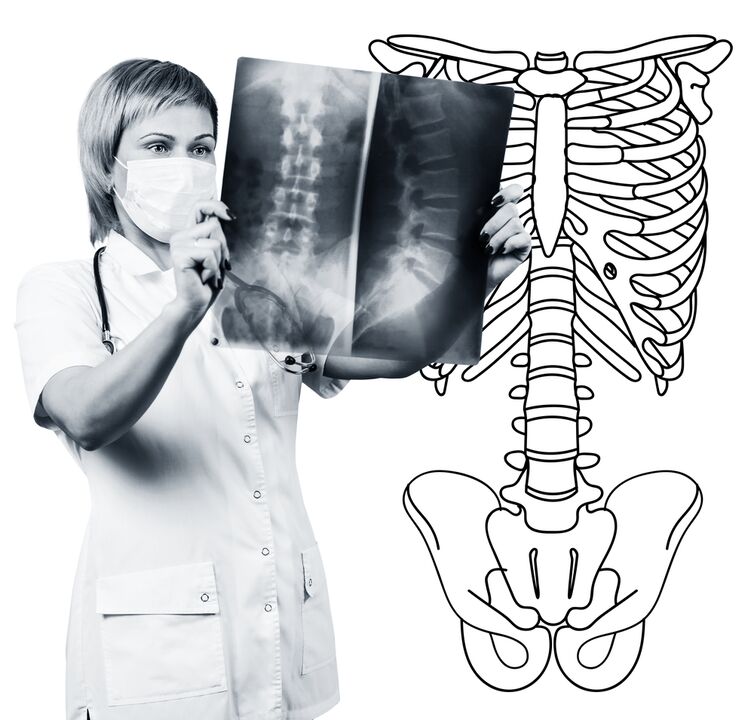
The diagnosis of lumbar column osteochondrosis is based on clinical data and additional research methods.The key role belongs to methods such as:
- x -ray of the lumbar column;
- Tomodensitometry of the lumbar column;
- Magnetic resonance tomography of the lumbar column.
The x -ray of the lumbar column is necessarily carried out in 2 mutually perpendicular projections - the rear and the right side.Such images allow you to see the shape, the contours and the structure of the vertebral bodies, the height and the shape of the intervertebral discs, the anomalies of the spine and natural turns.To display intervertebral joints and intervertebral holes, radiograms are produced in oblique projections.To identify the pathological mobility of individual lumbar segments (which is a sign of osteochondrosis), an x-ray is carried out under the functional test conditions, that is to say in the flexion and extension of the spine.Normally, you can clearly see the change in the height of the intervertebral discs in the front or rear sections in accordance with the direction of the body inclination, with osteochondosis due to the functional block of one of the segments, the height of the disc does not change or when flexion or prolongation.With pathological mobility, moving the vertebrae forward or back is determined.The main signs of X-ray osteochondrosis include the narrowing of the intervertebral slit, pathological mobility and the displacement of vertebral bodies, the deposit of salts in the disk fabric (calcification), the formation of regional growth of vertebral bodies, the compaction of vertebrates on the border with the affected disk (subchondral sclerosis).The x -ray of the lumbar column is a research routine, which gradually loses its meaning in the context of the active implementation of new and more informative research methods (CT and MRI).The x -ray of the lumbar department is used today as a method of screening diagnostic.
The lumbar column with the lumbar column is also made using X-ray radiation, but the radial load on the body is much lower than with the X-ray.The study is carried out lying on the table of a special device - a computer tomograph, it is absolutely painless.The resulting images are processed using a computer and allow you to see many more structures than with the radiography of the spine.
MRI is a method in which electromagnetic radiation is used to create images.The study is also carried out in a position to sleep on the table, which calls on the Tomographer's room.MRI is harmless and painless.
CT scan or MRI of the lumbar column allow you to see all the structures of the spine, to carefully examine the intervertebral discs (and the jacket and the fibrous ring) and the intervertebral holes, the content of the vertebral canal.Even a slight projection of the intervertebral disc will not go unnoticed.These methods (in particular MRI) allow you to determine the direction of the disk hernia, if necessary, the degree of compression of the nerve roots, the spinal cord.Thus, these research methods are much more informative in the diagnosis of osteochondrosis of the lumbar column than radiography.In addition, they allow you to diagnose not only osteochondrosis, but also other diseases (tumors, circulatory disorders in the spinal cord, abscesses, congenital anomalies of the structure of the spine and spinal cord), which is important during the differential diagnosis of causes of pain in the back.
Osteochondrosis of the lumbar column is a disease that most often causes back pain.It is in fact the destruction of intervertebral discs.Due to osteochondrosis of the lumbar column, a person often loses a work capacity, because, in addition to pain, the disease can lead to a violation of the mobility of the spine, the inability to sit, stand up and walk.Symptoms of this disease are not specific and require additional research methods to accurately confirm the diagnosis.The most informative and most sure of modern methods of diagnosis of osteochondosis is the MRI of the spine.